What was my biggest obstacle?
When I started working on medical computing, there might at most be one very large computer tucked away in the billing department of a hospital. There was no clear career path in "medical computing". There was not even an undergraduate major in computer science, so instead I majored in mathematics. I was one of very few women in computing and medicine. I was cracking the fields of both computing and medicine at a time when there were few women in either area.
The computing machines were primitive. Using a computer meant going to a computer center to use a punch card machine to input up to eighty characters of data or code on a card, a tricky process because insert and delete were multi-step processes. The cards were then submitted to the center to be run on the computer; in about three days, the results could be picked up. It was very important to review your code carefully, so as to prevent errors and reduce turnaround time to completion. A very good programmer did not make mistakes.
It took some nerve to see these hulking machines as the future in medicine. I had to search out any kind of opportunity I could find for training. My first summer job was at the National Biomedical Research Foundation where I used a plotter to draw tooth eruption charts. Later, I got a summer job at the National Bureau of Standards working with a small group that were putting the MUMPS computer language through the standardization process. These put me in contact with a number of key individuals involved in the foundation of medical computing.
I discovered there were a small number of medical schools and institutions that offered medical computing rotations for medical students. I spent three months at the National Institutes of Health, under Dr. Arnold Pratt, talking with a large number of individuals about what they did in medical computing. I spent another three month rotation at Harvard Medical School with Drs. Otto Barnett, Howard Bleich, and Warner Slack, pioneers in medical computing using MUMPS.
However there was still no advanced training in medical computing. I ultimately decided to do residency training in clinical pathology, which includes laboratory areas like hematology, chemistry, microbiology, and blood bank. I completed my residency at the Johns Hopkins University where I worked with Dr. Robert E. Miller on laboratory information systems.
At all times in my career, I have been in on the ground floor of Medical Infomatics. The Symposium on Computer Applications in Medical Care was just being formed in 1978. It provided an excellent forum on medical computing. I was one of the founding members of the organization and served the bylaws committee along with my husband, Vincent Brannigan, who acted as counsel. He became interested in the medical computing issues himself, and developed the area of medical privacy and liability.
There still was no clear career path. I was awarded a career development grant by the Dept. of Veterans Affairs to explore image analysis in pathology, looking at applications for platelet aggregation, muscle pathology identification and neuronal particle analysis. At the same time, I was elected to serve as Executive Director of the MUMPS Users Group (MUG). The MUMPS computer language was just beginning to be used in the VA for hospital information system functions. There were a number of international users of MUMPS, and I soon met Drs. Wolfgang Giere and Ichiro Wakai, my counterparts in MUG-Europe and MUG-Japan, who were extremely helpful in teaching and supporting me in this new environment.
My next position was with Georgetown University doing medical image analysis. I used a specialized image analysis computer. During this time, images were stored on 5mb disks that were approximately 12" in diameter. About 20 images could fit on one disk. The disks were labeled by hand in order to find the images later. It occurred to me that if the database technology that was so simple in MUMPS could be brought together with image storage technology, it would be tremendously helpful. However, technology was not ready for this until a few years later when PCs became more practical.
In the mid 1980's, I went on to integrate medical images with clinical database systems. This work was demonstrated at the VA Medical Center in Washington DC, and its potential was apparent to Dr. Ross Fletcher and Dan Maloney. I built a prototype system, integrating image capture and display capability with the VA's hospital information system. We began demonstrating this system within the VA, and clinicians were astonished and excited by the enormous impact it could have on medical care. The VA decided to fund a project to implement the system at a pilot site, the Washington DC VA Medical Center.
The biggest obstacle has always been the same one faced by any pathbreaker. It is not enough to do a job well, you have to recognize that the job needs to be done and convince others that it is important to do it. Many people with insight and imagination were critical to my success.
How do I make a difference?
I make a difference through my work in developing a multimedia patient record that can provide integrated information to physicians quickly and efficiently so medical decisions can be made optimally. Medicine is an information intensive profession, and this system changes the way medicine is practiced.
Prior to the initiation of this project, imaging systems were separate from database systems. Text and images were very different types of data with different technical needs in terms of management. Imaging technology and hospital information system (HIS) software technology were integrated by this project, for the first time. This combination provided far more functionality than the components could separately. The availability of information technology, advances in workstation displays, network capability, and mass storage devices were critical to the development of this application.
The VistA Imaging System joined with the VA's computerized patient record system to produce a complete Multimedia Patient Record that includes traditional medical chart information as well as a wide variety of medical images from specialties such as cardiology, pulmonary and gastrointestinal medicine, pathology, radiology, hematology, and nuclear medicine. Clinicians can place orders, write progress notes, and capture images online. They can access all of their patients' data quickly and conveniently from any clinical workstation.
The VistA Imaging System also allows radiologists to interpret images directly from high resolution workstations. They can get more information from an x-ray or other study because they can manipulate it to enhance different structures. Some find they tire less quickly reading from a workstation. Others feel more confident about their interpretations. Because of the enhancement capabilities, studies can be read that would otherwise require another exposure of the patient if film were used. Patients spend less time in the waiting room, and clinicians spend less time looking for x-rays.
The VA's Integrated Medical Imaging System has proved extremely helpful to treating clinicians, and saves them significant time. Physicians using the system have suggested new applications and a wider variety of data to be handled. It is used routinely when physicians meet together to discuss patients' diagnoses and plan their treatment courses. One physician user said "There's not a single GI case we do these days without the Imaging System. It makes a tremendous difference. We know what's there, like the tumor size. We can plan the operation. We can decide not to operate if it's a marginal case." The system allows easy, rapid and immediate storage of image information, often at the time of the procedure. This is important in saving physicians time and also because a patient's lesions may disappear or change quickly. Images are continuously available to multiple users at a time. The system makes communication between physicians more objective. Clinicians say "images provide more and better information than reports because it is so difficult to describe visual images in written form." A picture really is worth more than a thousand words.
The system helps VA physicians provide better care for their patients because decisions are based on a more complete picture of the patient's condition. The need to repeat procedures is greatly reduced. Images can be viewed simultaneously for treatment reviews and conferences. Images are available for immediate viewing at times when patient decisions must be made rapidly. Second opinions and consultations by physicians at other sites are possible using transmitted images and text. Physicians are better trained because the system's images provide a form of online continuing medical education. The VA's state-of-the-art information technology makes it easier to recruit good physicians. In addition, the system can be used to show patients their condition and allow them to make a better decision regarding their own treatment.
The VistA Imaging System was created to meet the needs of physicians in treating patients. Hospitals typically have an unsolved problem of handling the many and varied images used in patient care. These are stored in many forms, including xray films, glass microscopic slides, and video tapes, which are bulky, exist in only one copy and often suffer loss or deterioration. Treating physicians need comprehensive patient data including images and text to provide patients with high quality care. Information must be delivered in a user-friendly, straight-forward and simple-to-learn manner so that clinicians can get their data quickly without losing valuable patient-care time. This project combined existing information technologies to meet these needs with an emphasis on integration of functionality.
The VA's integrated imaging system has had an impact on the work patterns and thinking processes of physicians and has attracted worldwide recognition. In the past, clinicians have tended to be reluctant to use information technology. The VA's Integrated Imaging System is so clearly beneficial to medical care that it has overcome the traditional reluctance on the part of physicians to use computers. The availability of images combined with report data meets a need and has made them eager to use the technology. Information resource staff at V.A. medical centers also recognize the potential impact on medical care and have shown enormous enthusiasm and dedication in the setting up equipment for their users. They know that this system integrates with their existing information technology and will fit with the VA care providers way of doing business.
The integrated imaging system helps break down barriers between medical specialties by making all images available to every clinician, regardless of specialty. Recently, there has been an increased emphasis on the use of primary care providers. The multimedia medical record enables these providers by giving them access to all data needed to treat their patients. The technology also breaks down distance barriers because all patient information is available to the provider, independent of their physical location.
This project has shown that it is possible to build powerful efficient medically sophisticated hospital information systems on small budgets with high impact on the health care.
I have also made a difference through my efforts to assist technology transfer. I began this work when I was an officer of the MUMPS Users Group, an organization dedicated to the sharing of software. People from all over the world ran MUMPS, and shared much valuable software. The VA was one of the largest donors of software, through the Freedom of Information Act.
Later, I participated in a collaboration between the VA and the National Cancer Institute of Egypt, which was funded by the United Nations and U.S. AID. The National Cancer Institute of Egypt adopted the VA's hospital information system, translating critical portions into Arabic. Several other Egyptian hospitals later took the same approach. At the present time, the VA's computerized patient record system is operating in the NCI-Egypt.
This technology transfer work was important in bringing the benefits of computer technology to other countries. Simple functions like patient registration and scheduling can help enormously by allowing tracking of patients and efficient utilization of resources. More sophisticated software modules allow monitoring of disease and epidemiologic research.
Who was my mentor?
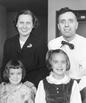
When people say "it takes a village" to raise a child, many of the village serve as mentors. I had a number of mentors along the way, some playing larger or smaller roles. However, my first and most important mentor was my mother, Dr. Margaret Oakley Dayhoff. My mother was a brilliant scientist, and the first woman to be secretary president of the Biophysical Society. She had fought a number of battles in establishing herself as a scientist, in a time when scientific institutions had rules like "no women can be full professors" or "we don't hire women as senior research scientists". She knew it was important that I get the experience and confidence necessary to succeed. My mother combined her extraordinary talents in mathematics and chemistry to create and mold a field that is now called Bio-informatics.
My mother recognized my interest in mathematics and science early, and guided my development in subtle ways. Throughout my elementary school years, she found special classes and activities to help me develop. On Saturday mornings, she took me with her to the computer science center at the University of Maryland. I quickly found that I could create pictures with letters punched onto cards and then printed on the computer printer. She found a programmed learning book on computers which I eagerly read. And she called my attention to an article by Claude Shannon on Self Learning Machines. Following the instructions, I built a self learning machine and demonstrated it to my junior high school class. The concepts in this article were a precursor to many of the ideas about artificial intelligence and neural networks, and it was very important to my development. My mother also led by example. She was very excited by her own work, and talked about it daily at home. She shared the good and the bad experiences, as well as the scientific details. There were many days when I would go to work with my mother - holidays, sick days, summer days. I often kept myself busy with school work. But sometimes, my mother would give my sister and I a job to do. One memorable task was punching out circular cellophane dots to build 3-D chemical models. Occasionally, I went on business trips or attended scientific meetings with her. From these experiences, I developed an internal understanding of the work environment, the importance of interactions with co-workers, and the kinds of activities that are expected. I knew that I would have a career when I grew up. This was in marked contrast to other girls in my generation who didn't discuss careers and weren't encouraged to think beyond college.
When I reached high school, my mother arranged a summer position at the National Biomedical Research Foundation where I would learn computer programming and work for colleagues in the separate computer building. I was assigned to a project developing programs to plot tooth eruption charts. Working in a punched card environment, I developed a method to draw each tooth shape in the correct position using a Calcomp plotter. In my spare time, I developed artistic computer drawings with a friend, and we won honorable mention in the 197 1 Computers and Automation Computer Graphics contest.
My mother's focus was developing statistical methods for comparing protein and nucleic acid sequences, a field that had been included in the broad domain of Biophysics. Early in her work, my mother saw the value of a database of sequence information that would allow researchers to build on each other's work. While in medical school, I co-authored a paper and a chapter in my mother's book, The Atlas of Protein Sequence and Structure, describing a new way to measure how closely proteins are related. She helped me prepare for my first scientific presentation.
In the 196Os, protein sequencing had opened up a new, objective way to look back over time at the evolution of living things. It showed the incredible similarities among the chemical structures that all living things are made of, and it could even help determine the shapes of proteins. Later in the 1970's, my mother began looking at nucleic acids using similar statistical techniques. At this point, the medical and pharmaceutical communities became very interested in this work because they recognized that sequences and the information that they provided would be important in the treatment of disease. However, it wasn't until recently that the field was named Bio-informatics, and has become a focus of Medical Informatics.
My father, Dr. Edward Dayhoff, was also important in my development. He was an experimental physicist who worked with magnetic resonance and with lasers. At home, he enjoyed building electronic devices in our basement workshop. He encouraged my interests in hardware, helping me build electronics projects when I was young. In the later 1970's, he became interested in computers himself, and started assembling them in the basement. He started with early Intel processors, homemade papertape readers, and a homemade chassis and keyboard. In 1980, he helped me put together an S-100 bus PC for my own use. Beyond my mother, I have had the good fortune to work with many gifted people who have helped me develop and find a path to accomplish what I set out to do.